Pleural thickening is a common finding on routine chest X-rays. It typically involves the apex of the lung, which is called 'pulmonary apical cap'. On chest X-rays, the apical cap is an irregular density located at the extreme apex and is less than 5 mm in width [ 1 ]. In the early twentieth century, a pulmonary apical cap was thought to be.. Apical pleural thickening on chest X-ray means that there is a white area of thickening at the very top of the lungs on the chest X-ray. This is a common benign finding which is most commonly seen from scar tissue. This can be on both sides of the lung or a single side. The cause is usually unknown. The finding increases as we age, but can be.

Representative chest Xray image of pleural thickening. a. Chest Xray... Download Scientific
Apical Pleural Thickening Seen Bilaterally
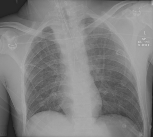
apical pleural cap pacs

“Must Know” chest RADIOGRAPH Radiology ppt download

Benign Pleural Thickening Radiology Key
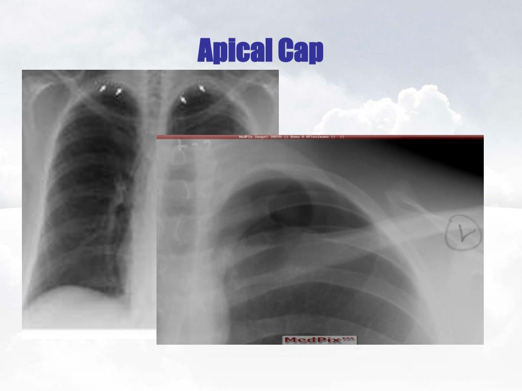
Icd 10 Code For Left Apical Pneumothorax
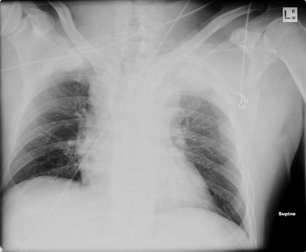
Pleurakuppenschwielen pacs

Apical pleural calcification Image

Apical Pleural Thickening

Southwest Journal of Pulmonary, Critical Care and Sleep Imaging Medical Image of the Week

Apical Pleural Thickening
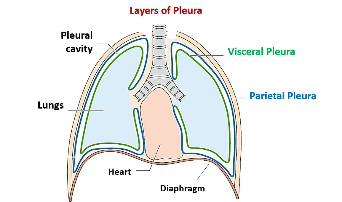
Pleura and Pleural Recesses Anatomy QA

bronchiectasis radiology apical pleural thickening ct scattered fibrosis in lungs CT Scan

Pleural Effusion On X Ray

Apical left extrapleural cap an early and important sign on chest radiographs Emergency

Matroană Pentru a căuta refugiu antic apical pleural cap pui de somn Cantină Cartofi

Chest Radiograph Showing Left Apical Pleural Cap Arrow Reconstructed Images

Apical View Chest X Ray

What Is Apical Pleural Capping
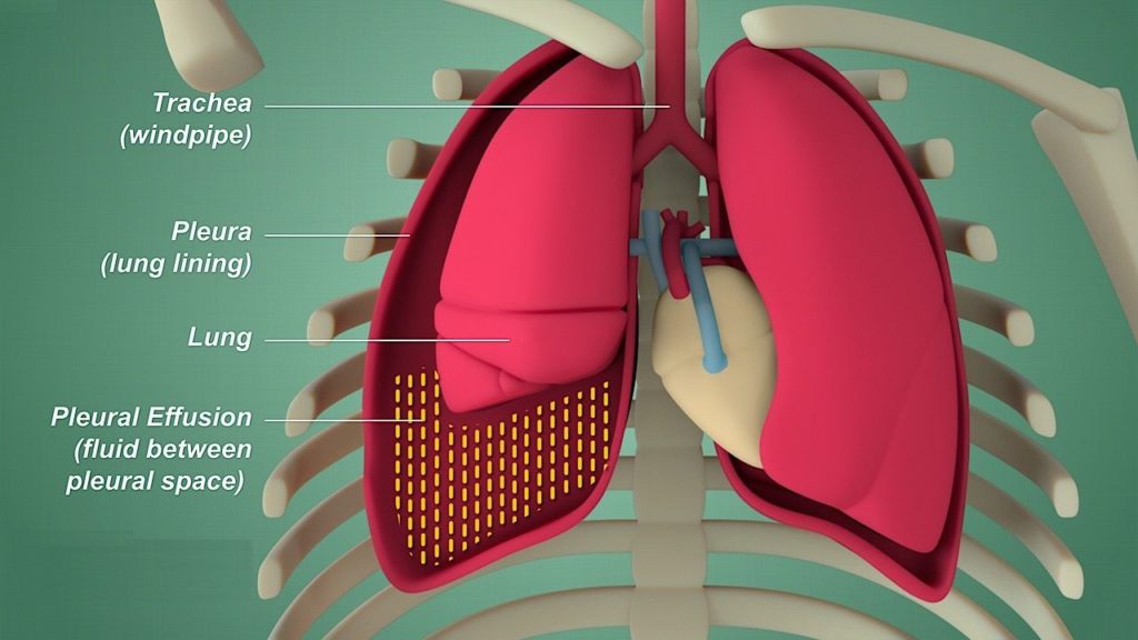
Wet, Wacky Lungs What is Pleural Effusion?
Apical pleural thickening (or "pleural cap") is often idiopathic and increases in frequency with age. If associated with prior tuberculosis, CT will demonstrate an increase in apical pleural fat. However, it is important to distinguish benign pleural capping from Pancoast tumour. Malignant thickening is usually thicker and asymmetrical, and.. Pleural thickening was found predominantly at the apex of the right lung. The apex of the lung was the most frequently affected area (Additional file 1: Table S2).Pleural thickening involving the apical area of either lung was defined as an apical cap, which accounted for 92.2% (n = 836/907) of the cases (Fig. 2a).More than half of the cases were bilateral and 35.7% involved thickening on the.