An x-ray of the ankle will have three views - AP, mortise, and lateral. It should be noted, though, that in some countries, including the UK, only the mortise and lateral are used.. by three months, 96.9% had returned to normal activities (it didn't matter which type of injury they had on MRI). Therefore, a removable brace may be.. A foot X-ray is a test that creates a black-and-white picture of the inside of your foot. The image displays the soft tissues and bones of your foot. These bones include your ankle bones (tarsal bones), the front end of your foot (metatarsal bones) and your toes (phalanges). A foot X-ray is also called a foot series or foot radiograph.

normal right foot x ray Google Search Foot x ray Pinterest Foot pain

Normal Left Ankle Xray

X Ray Of Foot

Normal foot xrays Image

Normal Left Ankle Xray
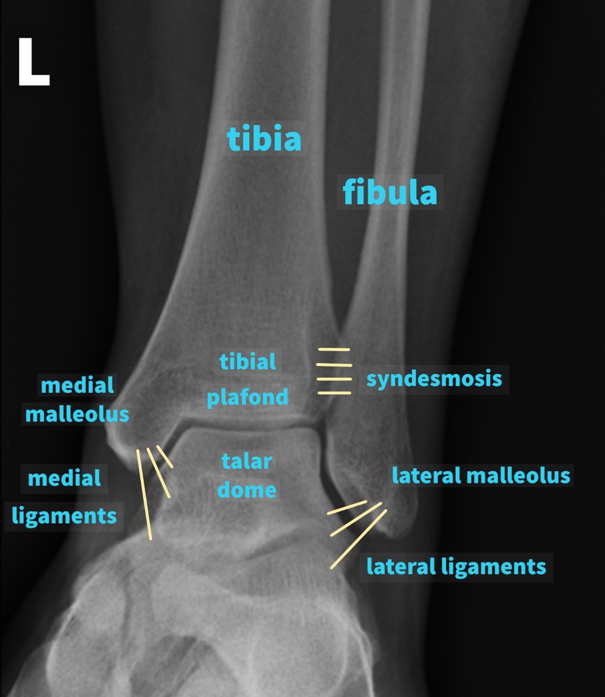
Ankle Xray Interpretation Ankle Fracture Geeky Medics

RiT radiology When to Obtain Ankle Radiographs
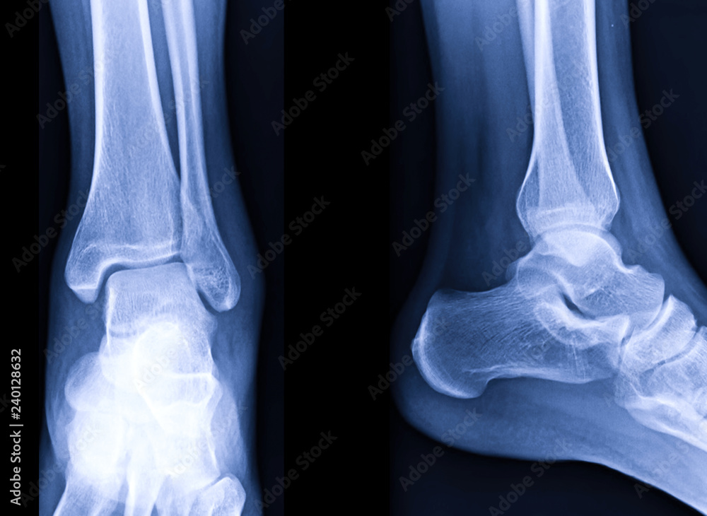
Radiographic image or xray image of Left ankle joint. Stock Photo Adobe Stock
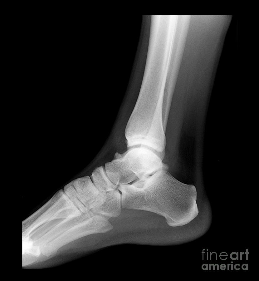
Ankle Xray, Normal Photograph by Living Art Enterprises

The Ankle

footxray Family Foot and Ankle

Approach to lateral radiograph of the ankle PULSE MD

Film xray normal child's ankle Stock Photo Alamy
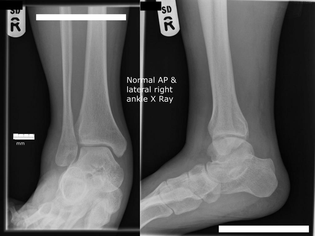
PPT XRay Rounds (Plain) Radiographic Evaluation of the Ankle PowerPoint Presentation ID

Normal ankle Image

Ankle X Ray Anatomy
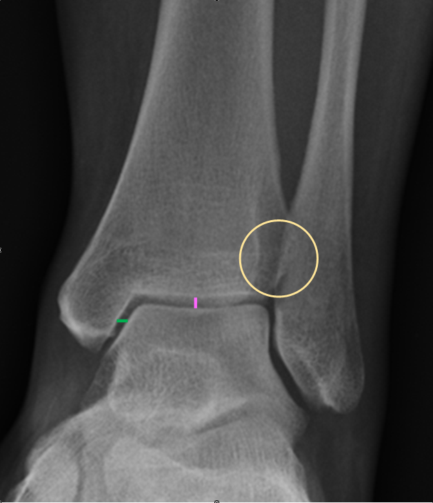
Ankle Xray Interpretation Ankle Fracture Geeky Medics

Normal foot xray ownnipod

Normal Left Ankle Xray
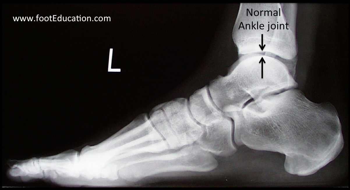
Ankle Arthritis FootEducation
A normal ankle x-ray reveals a symmetrical and well-aligned joint with distinct bone structures. The ankle consists of three main bones: the tibia, fibula, and talus. On an x-ray, these bones should appear smooth, with clearly defined contours and no evidence of fractures, cracks, or abnormalities. The tibia and fibula form the bony framework..